

About
About
The Confocal Microscopy Laboratory acquires and analyzes the fine structures of fluorescently labeled cells and tissues as high-resolution, three-dimensional images.
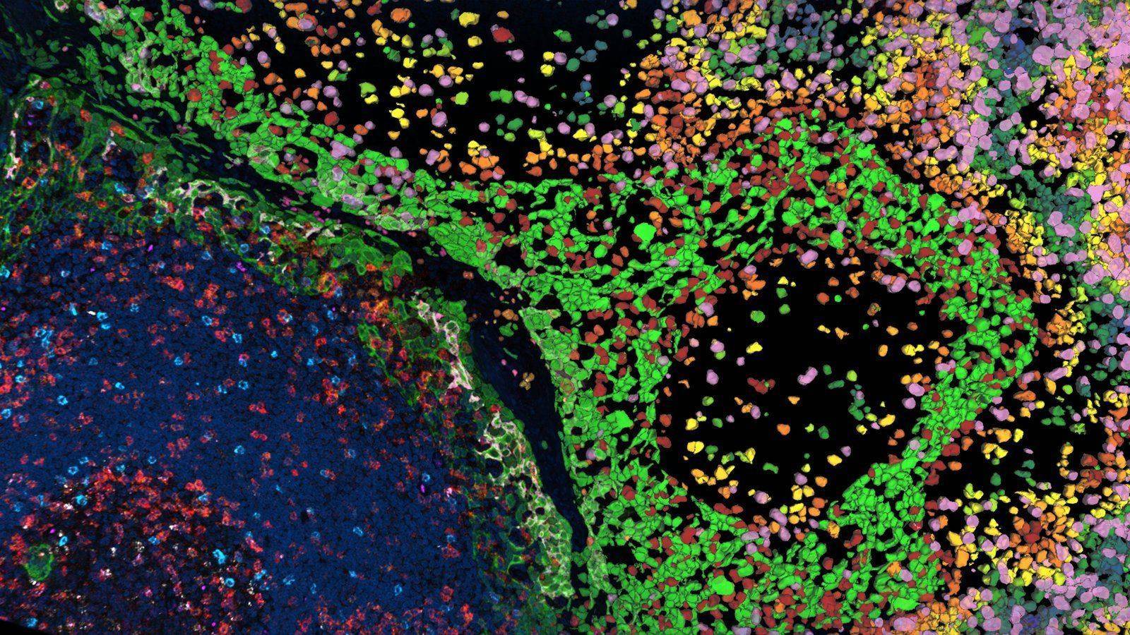
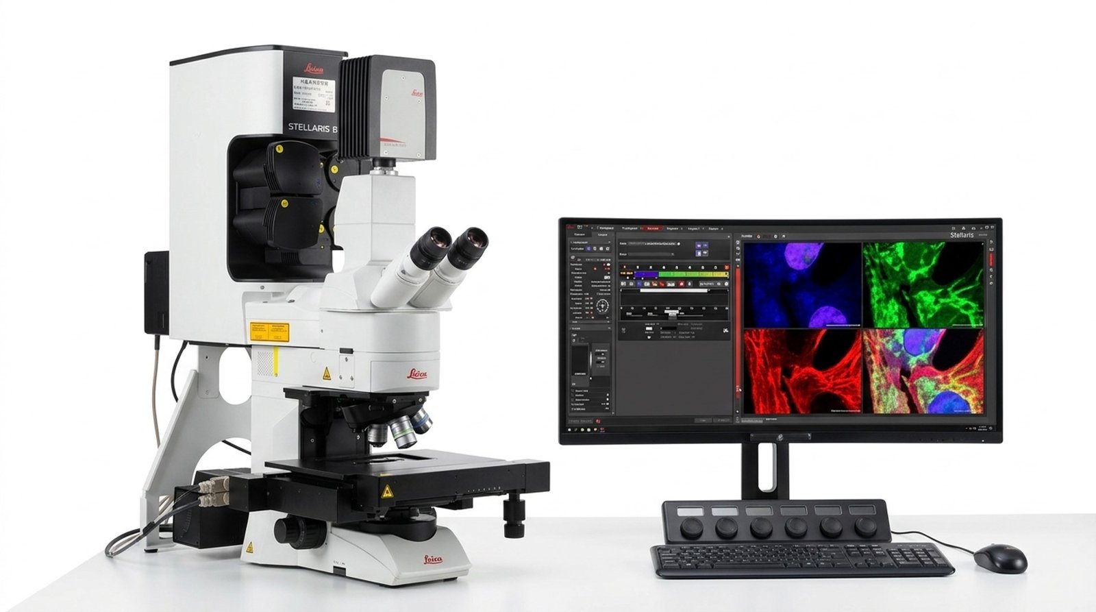
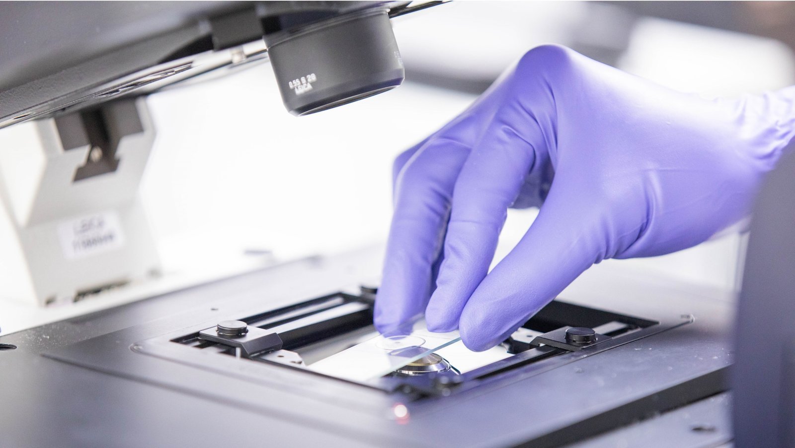
The Confocal Microscopy Laboratory uses Confocal Laser Scanning Microscopy (CLSM) to render optically sectioned specimens as sharp, accurate, high-resolution optical images.
It can be used for imaging and analysis of structures and biomolecules in both fixed and live specimens across diverse biomedical fields such as cell biology and immunology. The laboratory supports magnifications from 10x to 100x and high-speed scanning to image single- or multi-labeled specimens, and in particular enables 3D imaging, live-cell imaging, wide-field imaging using the Navigator function, and multi-fluorescence imaging.
The laboratory's equipment includes two inverted microscopes and one upright microscope, along with a Tissue Clearing System capable of clearing tissues or organoids. It is equipped with the LAS-X and IMARIS image analysis software, and in June 2026 the AI analysis program AiVIA was introduced. The laboratory also conducts regular training for individual users.