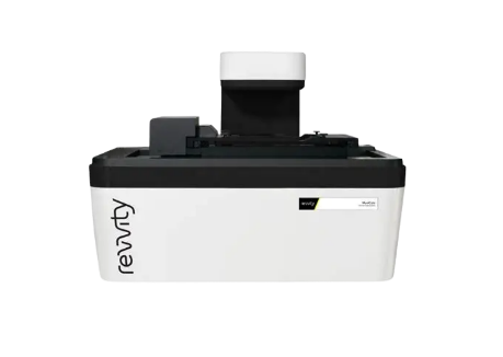
High Content Screening System - Operetta CLS
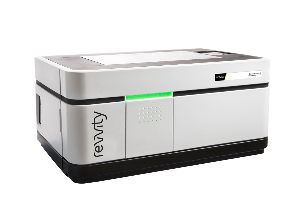
Operetta CLS High Content Screening
High Content Screening System Operetta CLS specifications
ModelOperetta CLS
ManufacturerRevvity (PerkinElmer)
Year Introduced2017
Usage TypeAnalysis Request (paid)
Features
  • Spinning disk type confocal
  • Automated imaging based on multi-well (96/348) plates (laser autofocusing) → analysis of characteristics at the individual cell level → quantification (phenotype classification)
  • Machine learning (PhenoLOGIC ML-based analysis) → coding-free analysis via the built-in analysis software → segmentation, phenotype classification, cell population, feature extraction, 3D culture, spheroid, organoid, and cell-surface texture analysis (image based)
  • Multi-imaging modality
  • Fluorescence, brightfield, digital phase contrast (DPC), and confocal (spinning disk) imaging available
  • Temperature and CO2 control available for long-term imaging
Usage
  • Reservation required 3 days before use → equipment warming up
  • Use of PhenoPlate (standard) recommended
LocationMedical Research Innovation Center (CMI), Room B2213

Real-time Cell Metabolism Analyzer - Seahorse XFe96
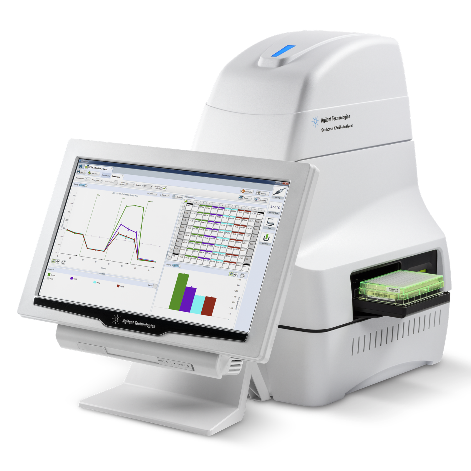
Seahorse XFe96 real-time cell metabolism analyzer
Real-time Cell Metabolism Analyzer Seahorse XFe96 specifications
ModelSeahorse XFe96
ManufacturerAgilent
Year Introduced2020
Usage TypeAnalysis Request (paid)
Features
  • Real-time kinetic analysis of mitochondrial respiration and cellular glycolysis via OCR/ECAR measurement
  • Label-free, non-invasive measurement (maintaining the live-cell state)
  • 96-well high-throughput (drug screening)
  • Automated drug injection system
UsageReservation required 1 week before use → equipment warming up; preparation of sensor plate and HCO3- free media; a cell pretreatment plan is required (prior consultation needed)
LocationMedical Research Innovation Center (CMI), Room B2213

Exosome Characterization System - ExoView R100
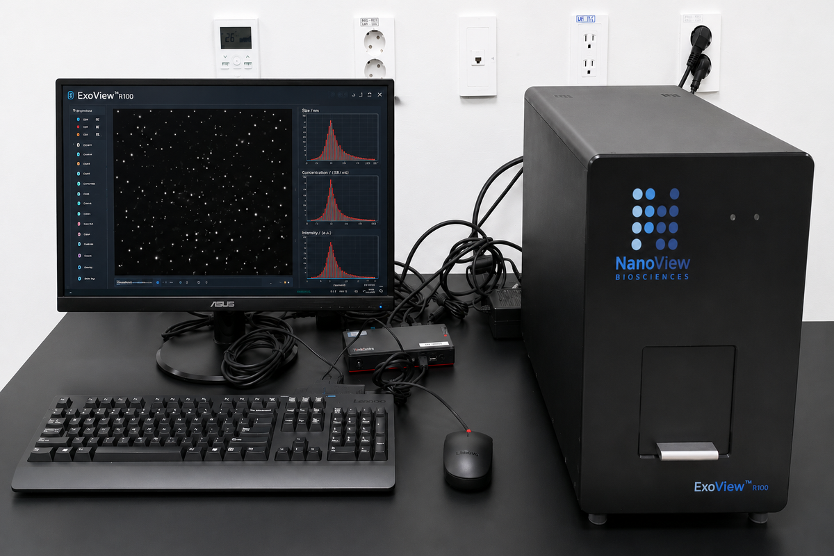
ExoView R100 exosome characterization system
Exosome Characterization System ExoView R100 specifications
ModelExoView R100
ManufacturerUnchained Labs
Year Introduced2021
Usage TypeAnalysis Request (paid)
Features
  • Particle size analysis (label-free method using interferometric imaging)
  • Exosome marker analysis (CD9 / CD63 / CD81 / exosome cargo via fluorescence labeling)
  • Single EV analysis → single-vesicle-level analysis rather than bulk EV analysis → heterogeneity analysis possible
  • Antibody-based capture (microarray chip) → direct analysis without EV purification (plasma, urine, CSF, cell culture media, etc.)
  • Multi-parameter analysis → particle count, size distribution, surface marker, co-localization, EV phenotype profiling
  • Applications: Cancer liquid biopsy, EV biomarker, drug delivery vesicle, immunology EV profiling
Usage
  • Reservation required 2 weeks before use → preparation time needed for dedicated microchip, buffer, washing solution, conjugated antibodies, and sample
  • Having prior extracellular vesicle counting data obtained with an NTA instrument helps optimize the sample loading amount
LocationMedical Research Innovation Center (CMI), Room B2213

Real-time Cell Imaging System - MuviCyte
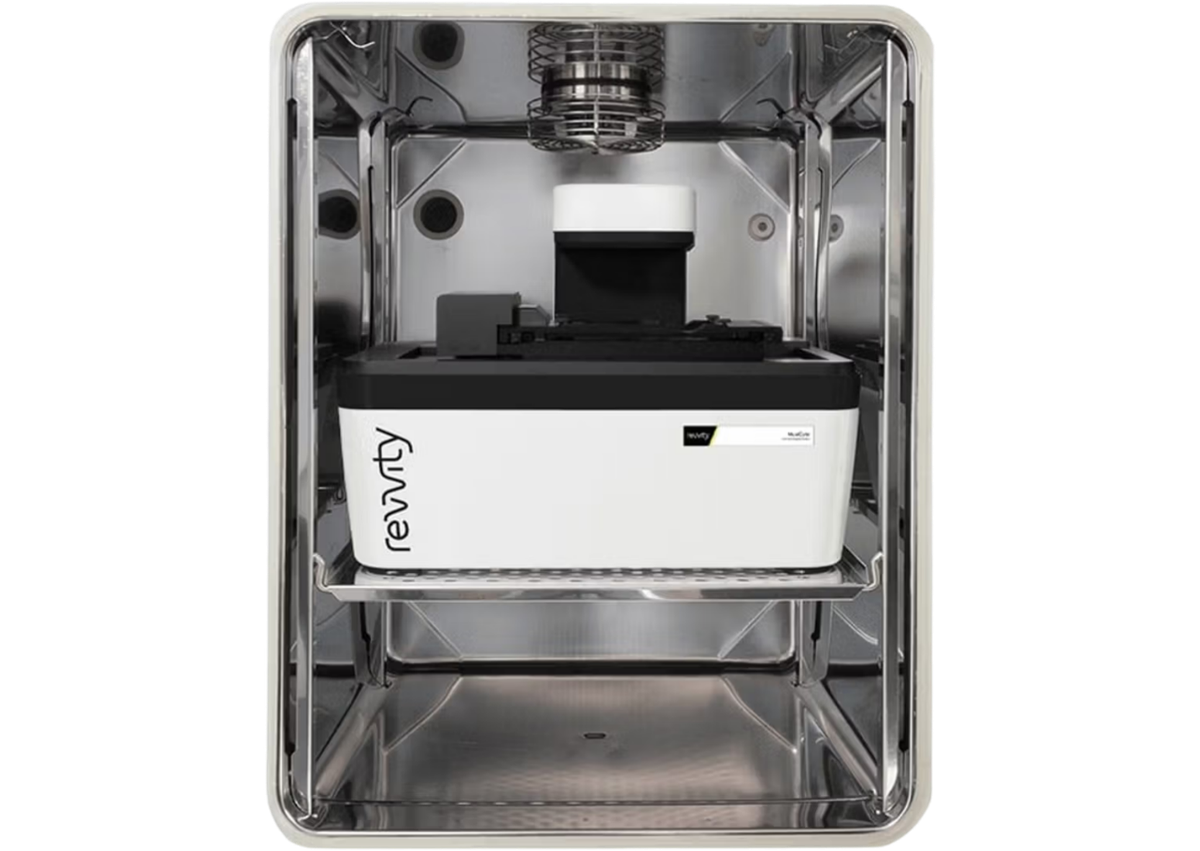
MuviCyte interior installation
MuviCyte exterior
Real-time Cell Imaging System MuviCyte specifications
ModelMuviCyte
ManufacturerRevvity (PerkinElmer)
Year Introduced2022
Usage TypeAnalysis Request (paid)
Features
  • A microscope installed inside an incubator (CO2/O2 adjustable) → minimizes cell stress / enables long-term observation
  • Long-term time-lapse imaging → records the entire process of cellular change (proliferation/apoptosis kinetics analysis)
  • Equipped with a 96-well wound scratcher (wound healing/migration)
  • Drug response kinetics
  • Cytotoxic assay
  • Spheroid growth
  • Stem cell tracking
  • Transfection efficiency
Usage
  • Reservation required 3 days before use → equipment warming up, CO2/O2 condition adjustment
  • When using a 96-well wound maker, use of SPL products is recommended
  • Fluorescence phototoxicity (repeated imaging at short intervals → induces cell stress → minimize exposure / imaging interval adjustment needed. Phototoxicity: GFP
  • Imaging interval:
    • Proliferation → 30~60 min
    • Migration → 5~15 min
LocationMedical Research Innovation Center (CMI), Room B2213