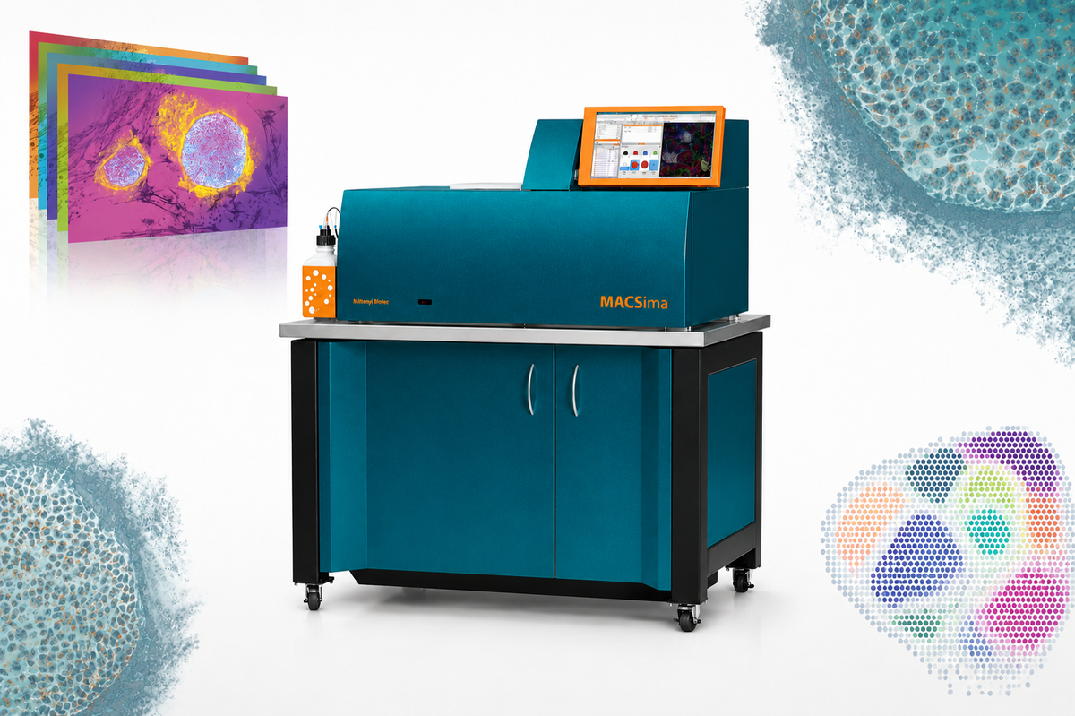
| Model | MACSima™ Platform |
|---|
| Manufacturer | Miltenyi Biotec (Germany) |
|---|
| Year Introduced | 2024 (first introduced in Korea) |
|---|
| Usage Type | Analysis request (paid) |
|---|
| Features |
- Cyclic multiplex imaging using MICS (MACSima Imaging Cyclic Staining) antibodies and erasing technology (antibody staining → image acquisition → fluorescence signal removal). Capable of analyzing 100+ markers in a single tissue (CMI has performed up to 64 protein targets). Spatial proteomics analysis.
- Segments tissue at single-cell resolution to obtain marker intensity for each cell. Analyzes not only fluorescence expression but also cell location (x, y coordinates) and relationships (cell-to-cell distance, microenvironment composition) - optimal for tumor microenvironment analysis.
- Fully Automated Workflow: The equipment automatically performs fluorescence staining (staining → imaging → signal removal → cycle repetition).
- Equipped with an Integrated Analysis Pipeline: Supports not only simple imaging but also data analysis (segmentation, feature extraction, gating, clustering UMAP/t-SNE, spatial analysis).
|
|---|
| How to Use |
- Reservation required 2 weeks before use (equipment warming up)
- Use of MACSima-standard imaging frame, buffer, and MICS antibodies is recommended
|
|---|
| Location | Medical Research Innovation Center (CMI), Room B2212-2 |
|---|